How is Stomach Cancer Diagnosed?
Endoscopy
Endoscopy is the most common way to diagnose stomach cancer. It can also be used to look for polyps, gastritis, ulcers and gastrointestinal bleeding. The procedure involves the insertion of an endoscope - a thin, flexible tube with a camera and light at one end that is guided through the mouth and down the gastrointestinal tract.
The patient is often sedated during the procedure to minimize discomfort. However the patient may choose to have a numbing throat spray with gargle instead of sedation. During the procedure, the doctor may decide to take a biopsy of the gastrointestinal tissue. A biopsy means that a small sample of cells is removed from the area of interest along the GI tract. This sample is evaluated in the lab for signs of cancer and other disease.
Video courtesy of Manipal Hospital.
IMAGING
CT (Computerized Tomography) scan is the most commonly used imaging tool. The CT scan is a series of x-rays from multiple angles. The images generated help the doctor see the inside of the body in 3-dimension.
In stomach cancer, the doctor may request a chest-abdomen-pelvic scan. The patient is often asked to drink a special contrast dye 1 hour before the scan. The dye helps to enhance contrast for a sharper image. The CT scan helps the doctor visualize the tumor in the stomach and the extent of spread to other areas of the body (metastasis).
Other imaging tests that may be used to detect the extent of stomach cancer include ultrasound, MRI and PET scan
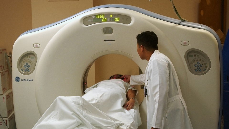
This image shows a CT Scan, which is a series of x-rays combined to create a single 3-D image of the body
LAPAROSCOPY
A laparoscopy is very similar to an endoscopy. The main difference is that a laparoscopy requires a small "key-hole" surgical incision (cut) through the skin of the abdomen. Laparoscopy is used to visualize the external surface of the stomach and surrounding abdominal cavity.
Endoscopic Ultrasound (EUS)
Endoscopic Ultrasound (EUS) combines endoscopy with an ultrasound. An endoscope is guided into the stomach. This time the endoscope has a small ultrasound probe at its tip. The probe emits high-frequency ultrasound waves inside the GI tract. The ultrasound waves bounce off the stomach and surrounding organs are are re-captured by the probe. The information is collected to create a 3-dimensional image of the location, size and depth of the tumor. A biopsy can also be taken during the endoscopic ultrasound procedure.
Biopsy
Three types of biopsies are used to diagnose and stage stomach cancer. An endoscopic biopsy takes a sample of the cells in the GI tract during an endoscopic procedure. A laparoscopic biopsy takes a sample of GI cells during a laparoscopic procedure. A fine needle biopsy (FNA) is done during an endoscopic ultrasound. A fine needle is inserted into the stomach wall, lymph nodes and surrounding organs and a sample of cells is collected. These cells taken during biopsy are then evaluated in the lab for signs of cancer. If cells are only detected in the stomach, the cancer is contained, however if more distal organs are positive for cancer, this means that the cancer has metastasized. See the next section for more information on staging stomach cancer.